Advanced Imaging
At One Heart Clinic we have access to the best quality advanced imaging equipment with fast turnaround times to ensure our patients have access to the care you need, when you need it.
Medical imaging is the technique and process of imaging the inside of your body for clinical analysis and medical intervention.
It gives us a visual representation of the function of organs or tissues in order to inform diagnoses and treatment plans for disease.
We use cardiac CT and MRI so we can see images of your heart, and how well it is working, including how your blood moves. The detailed, high-quality images produced by these scans, show your doctor your heart in two or three dimensions, to help us figure out what's going on, and whether there is a diagnosis to be made.
Your doctor may prefer one modality over the other based on your symptoms. Both scans are non-invasive outpatient procedures.
Advanced Computed Tomography (CT or CAT) scan uses X-rays — in many layered slices — to produce a detailed look at internal structures. Using this imaging technology in combination with an intravenous (IV) contrast (dye) helps clearly show the heart, vessels, and circulation. When the CT machine is used to look at the heart, it may be referred to as CTCA (CT Coronary Angiography), cardiac CT or CAT scan. All of these terms refer to the same test.
CT scans have no known side effects, and are usually pain-free. However, you will receive a low dose of radiation; if you have any questions or concerns about this, please get in touch. You will be carefully screened for suitability for the test and closely monitored throughout.
A cardiac CT scan is used to look inside the heart to fully evaluate the heart muscle, pulmonary veins, and coronary arteries. During this scan, images are taken from different angles and combined to produce clear cross-sectional images; this detail means the most minor abnormalities can be detected. The contrast dye also allows for a deeper non-invasive assessment.
Once in the scanning room, we will start an IV to administer the contrast solution while you’re lying on the scanner bed. This is normally put in a vein in your arm or hand. Electrodes that measure the electrical activity of the heart will also be attached to your chest. You will raise your arms over your head for the test, whilst remaining still and following breathing instructions from the team so that we can capture the clearest images.
After the test, you can continue with your regular daily activities.
To evaluate your risk for heart disease, your doctor may recommend a CT scan that provides a calcium score, which shows if there is coronary calcification in the arteries. If found, it can help predict future cardiovascular disease.
Those most at risk include those with a family history of coronary artery disease (CAD), men over age 45 and women over age 55, current or previous smoker, those who are overweight, and have been diagnosed with high cholesterol, high blood pressure, or diabetes.
While this is an excellent test in predicting particular types of coronary artery disease, it is not definitive due to certain soft plaques (build up) not being visible using this technology.
The scan is synced with your heartbeat to look for calcium deposits within the arteries. Once the test is finished, if calcium is noted, it will be given a score that estimates the extent of coronary artery disease.
Your doctor can discuss steps to take to lower your risk of developing heart disease. This test is normally performed under the guidance of a doctor who has specialist training in cardiovascular imaging.
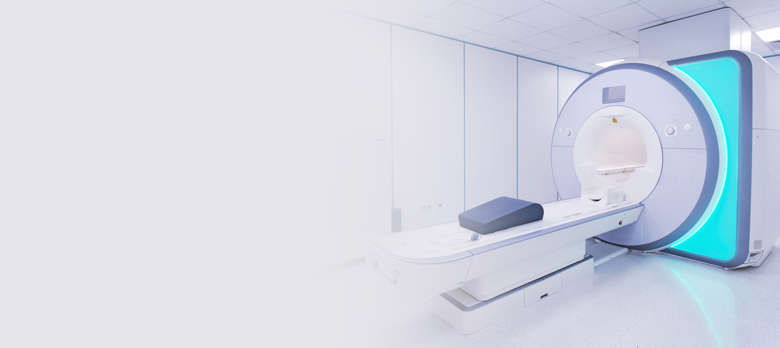
Magnetic resonance imaging (MRI) is a type of scan that takes detailed pictures of the inside of your body, whereas Cardiac MRI scan, is used specifically to provide a detailed image of the heart structure and composition of the heart tissue. These detailed, high-quality images in two or three dimensions help your healthcare provider figure out what’s wrong and make a diagnosis. Cardiac MRI scans also look at the blood supply to your heart.
Your doctor will order a cardiac MRI when they’re trying to diagnose a problem with your heart, such as the cause of your chest pain, shortness of breath or fainting, an enlarged heart, thickening of heart muscle, heart failure, heart valve disease and heart muscle damage, inflammation, and infection, amongst many other things.
Your doctor may even want to perform a Stress cardiac MRI, in which we will administer a drug intravenously (IV), usually in the arm of hand, so that we can observe your heart when it is mimicking the effect of exercise, whilst you are lying down in the scanner.
It does not use radiation, but if you have any questions or concerns, please get in touch with our team.
These images allow doctors to assess cardiac function, heart muscle scarring, and a potential lack of blood supply (which is known as myocardial ischaemia). CMR scans are considered the gold standard imaging modality for patients with cardiomyopathy as it allows for better heart visualisation, and is the only way to detect the heart muscle scarring, often seen in these conditions.
If your doctor has opted for the Stress protocol for this scan, it is to see whether the heart is being deprived of any blood supply when under stress. If this were the case, it may suggest blockages in the coronary arteries that require treatment. It is a safe test; in rare cases, a small number of patients have experienced a reaction to the drug, but you will be carefully screened for your suitability to have the test and closely monitored throughout.
During the scan, you’ll need to lie down on a long platform that will slide into the empty space in the middle of the MRI machine. We may put stickers with electrocardiogram leads on your chest and a belt below your chest to collect information about your heartbeats and breathing during your cardiac MRI scan.
The machine is quite loud during the scan, but you’ll be able to communicate with the person operating the machine. They will give you some instructions on when to breathe or hold your breath.
After the test, you can continue with your regular daily activities.